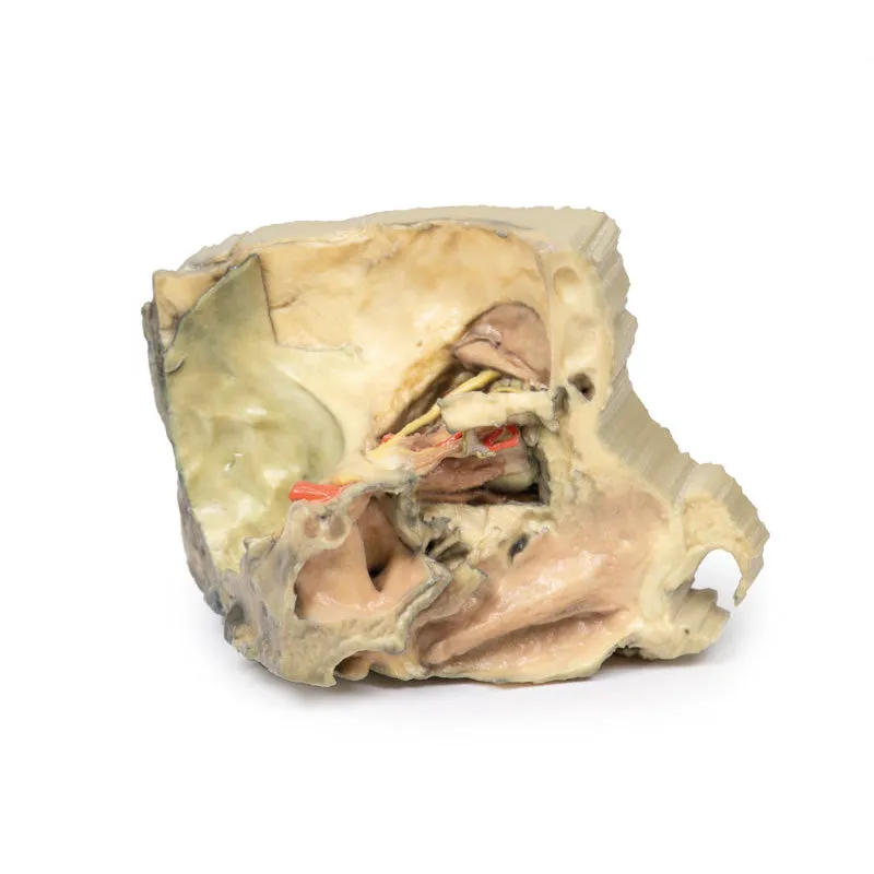
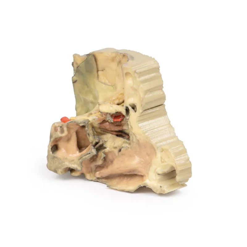
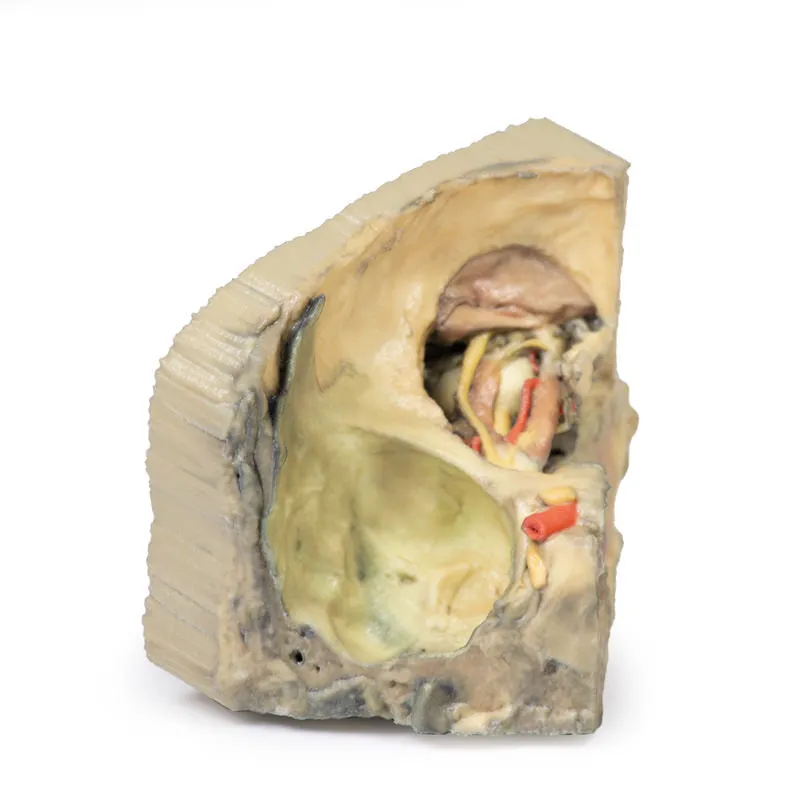
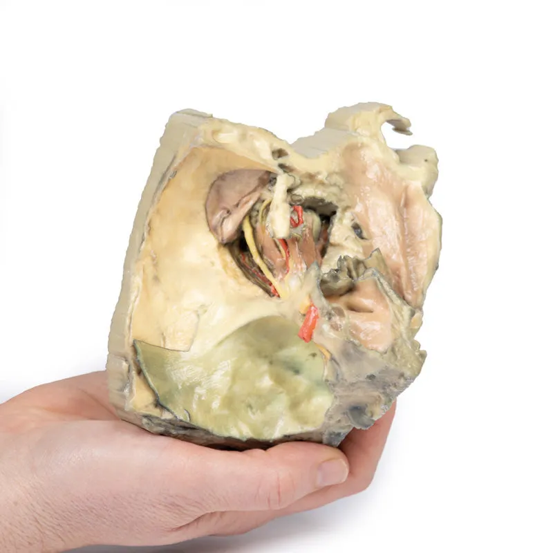
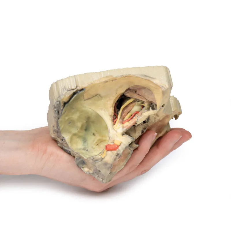
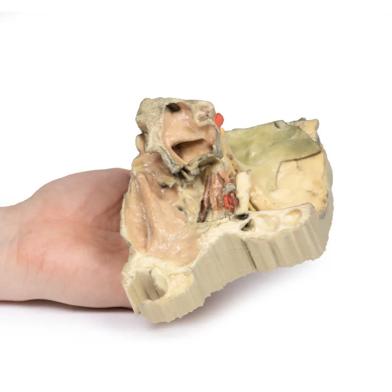
3D Printed Medial Orbit Replica
A small piece of the orbital plate of the ethmoid bone (EB) has been retained to illustrate its path as it enters the posterior ethmoidal foramen. Other structures visible include the frontal nerve (FN), the sphenoid sinus (SS), the pituitary gland (PG) and the frontal sinus mucosal lining exposed after removal of the orbital plate of the frontal bone on the anterior roof of the orbit. The internal carotid and optic nerve are also visible within the cranium.
Download Handling Guidelines for 3D Printed Models
GTSimulators by Global Technologies
Erler Zimmer Authorized Dealer

